tattoos

Wednesday, December 20, 2006

Ultrasound scan of rupture of uterus:


Rupture of the uterus during delivery is a known complication of pregnancy. The commonest cause is vaginal delivery after a caesarean section. Other causes include, previous surgery for tumors like fibroids and also prior classical caesareans, where the incision was placed in the upper uterus. Labor inducing medications can also cause rupture. I present ultrasound images of uterine rupture with severe hemorrhage on my ultrasound gallery at: http://drjoea.googlepages.com/ultrasoundgallery-uterus
On ultrasound scan, the typical findings are breach of the uterine wall, with a hypoechoic collection of blood outside the uterus. Follow up scans revealed diminishing of the size of the collection. One of the images of this case is shown here. Images courtesy of Dr. Durr-e-Sabih, Pakistan.

Monday, December 4, 2006

3-D ultrasound of animal fetuses:

Ever wondered how the fetus of an elephant, a dog or a dolphin appears on ultrasound?
Then try this link: Animal ultrasound
You dont need a diploma in radiology to identify the animal fetus :-)


Friday, December 1, 2006

The latest buzz on Teleradiology:

A recent survey by researchers in Yale University in New Haven, CT suggests that American physicians would prefer to have domestic radiology services, than having teleradiologists, based overseas, report the scans or X-rays of their patients. This survey reflects strong opposition, among a section of clinicians and radiologists, to the globalization of radiology services. While US based radiologists have a reason to feel threatened by their counterparts in Asia, because of loss of their share of the pie, clinicians are concerned about the quality of teleradiology services. More on this at the link:
Survey finds domestic teleradiology preferred by referrers
This represents insecurity and anxiety as the “dirty word” ie: “outsourcing” enters the US medical arena. But if banks, financial institutions and big businesses can do having their work outsourced to India, how long will it be before the American medics follow?
Dr. Joe Antony, MD.
Do visit:
free to view ultrasound image gallery>>http://drjoea.googlepages.com/
I also have a medical imaging blog at:http://www.healthvoices.com/taxonomy_menu/3/48

Thursday, November 23, 2006

The role of Ultrasound imaging in the diagnosis of scrotal swellings:


Swellings, with or without pain, of the scrotum, are very common in males. Swellings of the scrotum are commonly due to collection of fluid within the membranes covering the testes. These are called hydrocele. Other causes of swellings include testicular tumours (growths), which may be cancerous, a hernia descending into the scrotum, congenital conditions like an additional testes, cysts etc. The clinician has a hard time differentiating these various conditions, but a couple of minutes spent doing an ultrasound scan of the scrotum is sufficient to diagnose all these. I discuss the commonest cause of scrotal swelling, namely a hydrocele or pyocele. This condition can be easily treated by the urologist or general surgeon.
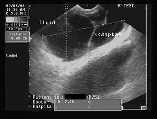
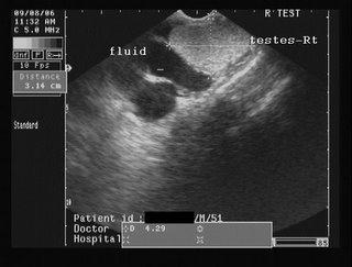
Hydrocele is an abnormal collection of fluid, usually serous, between the the two layers of the tunica vaginalis covering the testes. It may be congenital or acquired. Hydrocele is easily diagnosed by ultrasound scan. A pyocele is the same as a hydrocele, but has purulent fluid within it. Hematocele is a hydrocele with blood within the fluid and almost always is the result of trauma. Both pyoceles and hematoceles contain multiple septae (membranous partitions) within the fluid collection. At my web page: http://sites.google.com/site/drjoea/scrotum I present images of a septate collection, possibly a pyocele, and a large one at that, involving the right side of the scrotum.
DR. Joe Antony, MD.
A large free ultrasound image gallery can be found at:
http://sites.google.com/site/drjoea/home
Also visit: http://www.ultrasound-images.com/scrotum.htm
and http://www.ultrasound-images.com/acute-scrotum.htm for lots of information on sonography of scrotum.
For ultrasound study of scrotal infections visit:
http://www.ultrasound-images.com/scrotal-infections.htm

Case-2: moderately large right hydrocele/ pyocele with scrotal calculus:
This young adult male had a right testicular swelling of 3 months duration. Ultrasound images are shown below:


The ultrasound images show a large right hydrocele with particulate matter and septae. Possible infection of the hydrocele resulting in what is termed as pyocele.
Color Doppler image is shown below:


Color Doppler image shows almost normal vascularity in the right testis, ruling out orchitis.
Observe also the presence of the hyperechoic focus anteriorly, on the testicular surface- a scrotal calculus. This is literally a stone inside the scrotum, between the layers of the tunica vaginalis. Scrotal calculi can be mobile or adherent.
 

 This transverse section ultrasound video clip shows the magnitude of the problem, panning from the upper to the lower poles of the right testis.

Saturday, November 18, 2006

Sonographic diagnosis of conjoint twins:


Ultrasound Scan image (transverse section) of the conjoint twins sharing a common heart (such twins cannot survive). Images courtesy Dr. Durr-e-Sabih, Pakistan.


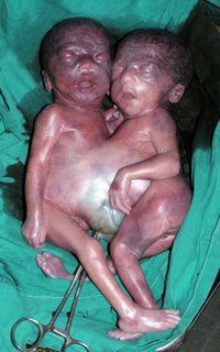
Post delivery view of the conjoint twins:
(below)


Conjoined twins or Siamese Twins (to the layman) are the result of incomplete division of the embryonic disc. The twins here are partially joint at either of the following points:
Head- called craniopagus
chest- thoracopagus
abdomen- omphalopagus
pelvis- ischiopagus
The incidence of such an anomaly occurring is very rare: 2 in 100000 births approximately. Fusion of the chest and abdomen is the commonest variety and is called thoraco-omphalopagus. It is associated with high mortality of the fetuses (most of the fetuses are born premature or still born). Ultrasound scan during pregnancy is very important to diagnose this condition.
Please click the link: http://drjoea.googlepages.com/obstetric-2
Here, I have presented ultrasound images of 2 different cases, with this anomaly.
Here, the twins are fused along the chest and abdomen with shared liver and heart. Such conjoined twins cannot survive. Diagnosis of conjoined twins is possible using ultrasound scan in the first trimester (first 3 months of pregnancy), but details are better visible in the 2nd trimester.
Dr. Joe Antony, MD.

free to view ultrasound image gallery>> http://drjoea.googlepages.com/

http://www.squidoo.com/conjoined_twins/
Also check my digg stories: http://digg.com/users/drjoea/dugg

Saturday, November 11, 2006

Role of ultrasound in ectopic pregnancy:

Sonographic diagnosis of ectopic pregnancy:
Ectopic pregnancy is a condition, wherein, the embryo (the baby in its earliest stage), is implanted outside the uterine cavity. The commonest location is inside the fallopian tube. As the fallopian tube cannot support the growing fetus, there is a danger of the sac (containing the embryo), rupturing with bleeding into the abdomen. Ectopic pregnancy is thus a potentially lethal condition. It should be suspected in any sexually active woman who complains of missed periods, pain in the lower abdomen (usually towards one side), and unexplained bleeding per vagina. Any such patient should immediately undergo serum -HCG and an ultrasound scan. Ultrasound diagnosis of ectopic is based on 1)absence of a gestation sac in the uterine cavity 2) presence of a gestation sac outside the uterus (usually to the right or left of the uterus ie: the adnexal regions). 3) The actual demonstration of a live embryo with heart beats is rarely possible. 4) There is usually a collection of free fluid or blood in the adjacent parts of the pelvis.
At my site: Diagnostic ultrasound gallery
I have described one such case wherein a live embryo of 7 weeks is seen in the right adnexal region. See page: http://drjoea.googlepages.com/obstetric-2
The high resolution images on the page leave no doubt about the diagnosis.Also see my blog at: http://drjoea.bloglanding.com/?method=comments&postid=12675#
Dr. Joe Antony, MD.

Friday, October 27, 2006

A calculus affair:

The mystery of the renal calculus:
Any sonologist worth his degree would have seen numerous renal calculi or kidney stones. But few would ponder over how many of the minute particles, yes particles, are actually calculi. The problem arises when one begins to diagnose stones of less than 4 mm. This is when the diligent radiologist or sonographer, faces the dilemma of labeling an echogenic structure in the kidneys, a stone or an otherwise normal appearance. Many bytes have been spent on the internet discussing these fine details with my colleagues. The hallmark of the renal or any calculus has always been the bright (echogenic) focus with a trailing acoustic shadow (a dark area behind the stone). As these pictures show, the shadow may not be obvious. In some cases, there may be a faint shadow, which further confuses the sonologist. These images by Dr. Ravi Kadasne, radiologist, in UAE, show a case where there is an echogenic lesion or focus followed by a faint shadow behind it. However, CT scan showed no such calcification or lesion. The best advise in these cases would be to do a follow up of the lesion after a few months.


Friday, October 20, 2006

Chikungunya update:

These bits of information may be useful to those afflicted by the illness:
1) As of 20th October 2006, there have been approximately 50,000 reported cases of this disease in Kerala.
2) It is spread by the Aedes Egypti mosquito, which bites primarily during the day; but in particular, during the dawn and dusk hours.
3) There is no proven effect of papaya extracts or juice as a remedy for Chikungunya. There have been many emails circulating this misinformation.
4) Pregnant ladies must be very cautious and seek medical help if suffering from Chikungunya, specially in the first 3 months (1st trimester).
5) There is no evidence of transmission of the Chikungunya virus via breast milk from mother to infant.
6) It generally takes upto 5 days for the serological tests (blood tests) to reveal the presence of the disease (positive tests).
7) Dengue lasts upto 2 to 3 weeks ; Chikungunya lasts upto 4 to 6 weeks. The joint pain may persist even longer, specially in the elderly patients.

Monday, October 9, 2006

DR. Joe Antony

Chikungunya hits Kerala:

A mosquito borne viral fever from East Africa has made it to India. Among the states affected are Rajasthan, Karnataka, Tamil Nadu and Kerala.
Chikungunya is a viral disease spread by the bite of the Aedes Aegypti mosquito, which breeds preferably in fresh water. How did this illness which was last heard of in the outbreak in Reunion island, spread to S. India? No one knows for sure. But it could be due to travel of infected patients from E. Africa. In any case, the Aedes Aegypti mosquito is no stranger to this part of the country. Lack of proper drainage of water is the primary cause for the flare up of this mosquito.
The symptoms of this disease are fever, rashes on the chest and limbs, and most important of all- severe joint pains. In fact, the joint pain and swelling (arthritis) is the hallmark of this ailment. The word Chikungunya means “which makes one bend” in Swahili (an African language). The patients prostrate and crouch due to the arthralgia (joint pain). Chikungunya is self limiting, though it can be fatal.
Treatment is symptomatic- use of antipyretics (for fever) and anti- inflammatory drugs for joint pain (arthralgia). Chloroquine phosphate is being tried in resistant cases.
In Kerala, nearly 100 deaths are attributed to this disease. But in the absence of detailed follow up and pathological studies, the true figures may remain hidden from the people.
The PCR (polymerase chain reaction) serological test remains the gold standard for identifying the disease. This has reportedly an accuracy of nearly 100 %.
Among the leading labs performing the PCR analysis in Cochin, include DDC- international and EI lab.
Chikungunya update: (12th Oct 2006)- 1.3 million cases reported across India.
Cases reported in Kerala- approximately 50,000.

Wednesday, September 20, 2006

Diagnostic Ultrasound Gallery

Interested in Ultrasound-- then visit our sister web site:
Diagnostic ultrasound gallery
You'll find loads of ultrasound images with a brief description of each case, links etc.

The crisis facing the small clinic in India:

The small, friendly neighbourhood clinic has for long been the backbone of medical practice in India. But of late, the arrival of large hospitals or multispeciality hospitals is rapidly changing the scenario.
In Cochin, where I work, many of the smaller and even midsized hospitals are feeling the heat, as large hospitals mushroom all over the place. The situation in the field of radiology is also undergoing rapid change. Until 5 years back, independent diagnostic centers ruled the turf of radiology. There were diagnostic centers with CT scan and ultrasound facilities “under one roof”. Then MRI scan was also added to the range of services. When even that was not sufficient, pathology labs were added to these “centers”. Even large hospitals would refer cases to these diagnostic labs. But, soon, these same hospitals decided that, rather than buy the cake, they might as well make it. The result, almost all the major hospitals in town have their own complete diagnostic facilities. The situation is so bad, that some of the independent labs have shut shop or shifted their equipment to greener pastures.
So, what does the future hold for medical practice here. Obviously, many of the numerous labs (and also the smaller hospitals) in this part of the world have to adapt to the changing times; many might simply close shop. Others would merge with the large mega-hospitals, to survive. The friendly neighbourhood clinic, or lab might operate on a part- time basis, with the radiologist operating part time at a number of labs.
The trends one sees in the corporate world will be mirrored in the medical field also.
Kerala, with its high literacy rates (almost 100%) and consequent health consciousness, coupled with inflow of petro- cash (from overseas Indians in the Arab countries), has an abundance of private healthcare facilities. In fact, it is estimated, that, Kerala has one of the highest number of CT scan machines, per unit population in the world. The changes in this microcosm will reflect those in the West and elsewhere.

Sunday, September 10, 2006

The future of MRI scan- Functional MRI:

Functional MRI scans – the future of MR imaging:
The latest in imaging has arrived, in the form of Functional Magnetic Resonance Imaging or F- MRI scans.
Imaging remained largely a means to study anatomical lesions of the body, such as tumours, infections etc. But now, F- MRI is set to change all that. F- MRI is a technique that actually maps hemodynamic changes (read blood flow changes) in response to
neural stimuli or neural activity, in the brain and spinal cord. This is based on the fact that that there are changes in blood flow patterns in regions of the brain that are active; for example moving the fingers would produce more blood flow and oxygenation in the region of the brain called the motor cortex. The MR signals of the blood vary according to the degree of oxygenation. This fact is made use of to color code the brain’s activity. F- MRI can thus non-invasively detect human brain tissue activity, without the fear of radiation.
I had the chance to see a slide show, where an F- MRI scan of a patient with anxiety disorder, was displayed. Multiple areas (colored red) were seen along both cerebral hemispheres. After making the same patient relax, listening to gentle music, a repeat F MRI scan showed a near normal appearance. Thus, the role of this technique, in neurology and psychiatry is obvious. A more controversial use of the technique in comatose patients, found that in some such cases, in so called minimally conscious states,
the brain showed responses to speech and external stimuli, similar to those in normal persons. This raises many questions, indeed a Pandora’s box of legal and ethical issues regarding the conventional definition of death.

Wednesday, August 30, 2006

HOW SAFE IS YOUR CT SCAN?

Risk of radiation during CT scans:
CT scans have become commonplace and are an essential part of investigations,
that the consultant/ doctor advises, today. However, have CT scans been taken for granted, as truly safe for the patient?

Recent studies done (http://www.fda.gov/cdrh/ct/risks.html ) in USA and Europe suggest that there is a small but significant risk of radiation induced cancer, occurring due to
CT scans. These conclusions have been made after the release of the BEIR VII report
( BEIR VII Press Release: Low Levels of Radiation May Cause Harm ).
According to the report, the National Academy of Sciences, has described a 1-in-1000 chance of developing cancer from a single radiation exposure of 10 mSv.
(mSv is a unit of measuring the amount of radiation a person receives).
10 mSv is the amount of radiation a patient receives during a single CT scan study of the
abdomen, chest or the pelvis. The study also finds that the risk in children is even
higher. 1 in every 550 children scanned would develop cancer due to the CT scan.
The recommendations of the study are that persons under 40 yrs. age should be carefully
screened to avoid unnecessary scans. This is specially true whilst scans involve the gonads.
A CT scan of the head exposes you to about 2 mSv, a scan of the chest is about 8 mSv, and a study of the abdomen and pelvis is about 10 mSv. A normal person is
exposed to 3 to 5 mSv of radiation every year (ie: radiation from soil and atmosphere).
The real problem arises when a person (especially those under 40 and children) are
subjected to repeated scans, with doses exceeding 50 mSv.

Conclusion: Be judicious in the use of CT scans. Though there are definite risks
involved in repeated scans, they are to be carefully weighed against the numerous diagnostic benefits this tool provides.

Monday, July 10, 2006

CLICK FOR FREE MUSIC


Looking for free music downloads, try this internationally acclaimed site:

Click Here To Download Free Music and Movies

Scan your computer hard drive for malicious software, adware, scumware etc.
you might have picked up from the internet.


Thursday, May 4, 2006

Dr.Anatoly--a radiologist and an artist


My good friend, Dr. Anatoly, from Kiev, Ukraine,
feels the human body and its insides
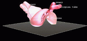
are a work of art to be perfected through 3D
graphics as he demonstrates so well in these pictures.
They show the female reproductive organs in
3Dimensions. Unfortunately, this site does not
allow moving 3D- animations which is what these are.

Sunday, April 23, 2006

AN AMAZING, TRUE, MEDICAL FAIRYTALE

Did you know that fairy tales could turn out to be as real as reality?
This is one that captures the imagination. It is woven around a background of war, intertwining the
destiny of strangers in far away lands. It is about friendship between people who have never met,
except through the medium of the internet. Strangers, who've never seen each other, except through
photographs on a computer monitor.
It is about a group of people - a doctor in Afghanistan, Dr. Wahaaj, and his struggles in war ravaged Afghanistan and a sonographer based in the USA, a lady named Marveen Craig.
The link: http://www.sonoworld.com/Sonoworld/Editorials/Marveen.aspx tells the story of how "determination
and perseverance can change lives". It is about how ordinary people can do extraordinary feats.
Why does this story concern me? I know both Wahaaj and Marveen, of course, in the virtual world.
We have talked, exchanged pictures, but never met. Both of them are dear friends, whom I have known
since the time I have joined MI, a email group devoted to medical imaging.

Friday, April 7, 2006

Really cool web sites- my selection

A) Business and finance:
1)FOR Indian MUTUAL FUNDS this is the best:
www.valueresearchonline.com
It has a lovely portfolio manager. I could not find any that can compare with this one.
2) For advise on ways to make money, especially, internet based small business-
this is good: www.seekluck.com
3) To check the sensex value and the share market: try http://www.bseindia.com/
or you could try the NSE at : http://www.nseindia.com/
B) about all things Cochin and Kerala: try this good blog- http://indulekhainfo.blogspot.com/
It covers current hot topics in Kerala, especially on the literary front, books, cooking etc.
It also has a detailed explanation on how to download and install Malayalam font.
C) For POP supported email services:1) try http://www.usermail.com/
POP or post-office protocol is what enables receiving email via Outlook, Eudora or other email
client software. Usermail charges a small fee, but has excellent service. I have been using it for
the past 3 years. It is virus free and spam free.
2) Gmail is also an excellent option. But the only problem is that you've got to be invited by a Gmail
member.
D) To help you choose that fancy digicams, TVs, PDA and mobile phones this
web site is great. It has prices and detailed specification of the latest and older models.
http://www.myproductadvisor.com/mpa/home.do
You can even compare the various features of all the models that catch your eye.
E) Want to verify whether that email or information you received is fact or fiction.
Try this site: http://www.truthorfiction.com
F) To translate this web-site to other languages, try this link:
http://babelfish.altavista.com/
G) For those magic home remedies for every ailment, try this site: http://www.mothernature.com/index.cfm
This site has many useful tips, which doctors themselves may find useful.

If you are interested in ultrasound, this web site is a must. It has a large collection of ultrasound images with a brief description of each case. Visit: free to view ultrasound gallery>>http://drjoea.googlepages.com/

Wednesday, March 29, 2006

Dr. Joe's ULTRASCAN CENTRE


Hi,

I am DR. JOE ANTONY, a radiologist having an ultrasound scan clinic here in Cochin, Kerala, S. India.
I propose to set up Cochin blogs as a site for guiding people to good hospitals,
diagnostic centres and nursing homes here in Cochin.
I also intend to set up pages (on this blog site) to explain sonographic techniques, images etc.,
for the medical professionals and the lay person.
My ultrasound scan clinic-- ULTRASCAN CENTRE is located at No. 34, LIG COLONY, JUDGES AVENUE, OPPOSITE SPENCERS, KALOOR, COCHIN- 682018. The location is ideally placed, just 2 Kms. from
the North Railway station (Ernakulam Town station) and has a City Bus stop just close to it.
Auto- rickshaws and taxis are easily available here.
Ultrascan centre is located just opposite SPENCERS shopping mall at Judges Avenue, Kaloor, Cochin.
And yes, there is car parking space available, close to my ultrasound clinic.
FACILITIES AT ULTRASCAN CENTRE:
1)My ultrasound scan centre uses the state of the art high resolution NEMIO XG (TOSHIBA) COLOR DOPPLER ULTRASOUND machine.
The ultrasound and color Doppler machine has 3 probes - a convex 3.5/ 5  Mhz abdominal probe for scan of the abdomen and pelvis; I also use a multifrequency high resolution endocavity probe for scan of the
female pelvic organs- uterus, ovaries, vagina etc.
The testes, thyroid and breast can also be studied with this probe.
In the male, I use this probe to scan the prostate by the transrectal route.
All these scans are done with a disposable protective sheath covering the probe.
For imaging of the breast, scrotum and thyroid etc., we use the high frequency 7 to 11 Mhz linear probe.
Almost every superficial part, including the joints and musculoskeletal regions can be scanned with this ultrasound probe. The color Doppler function of this small parts linear probe helps us to perform excellent color Doppler imaging (scans) of the carotid artery, jugular veins and veins and arteries of the limbs.
Varicose veins and deep vein thrombosis are also imaged well using the color Doppler system.
2)Broad band- Net connectivity and internet based consultation: any interesting or unusual cases
are transmitted via the internet to groups of sonologists/ radiologists world wide for immediate
exchange of views and ideas.

3) AMPLE SPACE: Ultrascan centre occupies nearly 1200 sq feet of space.
The patient's waiting room is large and can seat at least 20 people at a time.

4) The clinic is airconditioned. Back up power is available in case of power outage.
CONTACT ME at:
91- 484- 2323088 (res)
91- 484-2403058 (clinic)
9388623088 (mobile).
WORKING HOURS: 9:30 AM TO 5:00 PM. (IST).
Email:  drjoea (at) gmail (dot) com
 

blogger templates | Blogger