tattoos

Thursday, November 23, 2006

The role of Ultrasound imaging in the diagnosis of scrotal swellings:


Swellings, with or without pain, of the scrotum, are very common in males. Swellings of the scrotum are commonly due to collection of fluid within the membranes covering the testes. These are called hydrocele. Other causes of swellings include testicular tumours (growths), which may be cancerous, a hernia descending into the scrotum, congenital conditions like an additional testes, cysts etc. The clinician has a hard time differentiating these various conditions, but a couple of minutes spent doing an ultrasound scan of the scrotum is sufficient to diagnose all these. I discuss the commonest cause of scrotal swelling, namely a hydrocele or pyocele. This condition can be easily treated by the urologist or general surgeon.
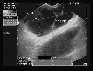
Hydrocele is an abnormal collection of fluid, usually serous, between the the two layers of the tunica vaginalis covering the testes. It may be congenital or acquired. Hydrocele is easily diagnosed by ultrasound scan. A pyocele is the same as a hydrocele, but has purulent fluid within it. Hematocele is a hydrocele with blood within the fluid and almost always is the result of trauma. Both pyoceles and hematoceles contain multiple septae (membranous partitions) within the fluid collection. At my web page: http://sites.google.com/site/drjoea/scrotum I present images of a septate collection, possibly a pyocele, and a large one at that, involving the right side of the scrotum.
DR. Joe Antony, MD.
A large free ultrasound image gallery can be found at:
http://sites.google.com/site/drjoea/home
Also visit: http://www.ultrasound-images.com/scrotum.htm
and http://www.ultrasound-images.com/acute-scrotum.htm for lots of information on sonography of scrotum.
For ultrasound study of scrotal infections visit:
http://www.ultrasound-images.com/scrotal-infections.htm

Case-2: moderately large right hydrocele/ pyocele with scrotal calculus:
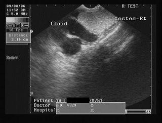
This young adult male had a right testicular swelling of 3 months duration. Ultrasound images are shown below:


The ultrasound images show a large right hydrocele with particulate matter and septae. Possible infection of the hydrocele resulting in what is termed as pyocele.
Color Doppler image is shown below:


Color Doppler image shows almost normal vascularity in the right testis, ruling out orchitis.
Observe also the presence of the hyperechoic focus anteriorly, on the testicular surface- a scrotal calculus. This is literally a stone inside the scrotum, between the layers of the tunica vaginalis. Scrotal calculi can be mobile or adherent.
 

 This transverse section ultrasound video clip shows the magnitude of the problem, panning from the upper to the lower poles of the right testis.

Saturday, November 18, 2006

Sonographic diagnosis of conjoint twins:


Ultrasound Scan image (transverse section) of the conjoint twins sharing a common heart (such twins cannot survive). Images courtesy Dr. Durr-e-Sabih, Pakistan.


Post delivery view of the conjoint twins:
(below)


Conjoined twins or Siamese Twins (to the layman) are the result of incomplete division of the embryonic disc. The twins here are partially joint at either of the following points:
Head- called craniopagus
chest- thoracopagus
abdomen- omphalopagus
pelvis- ischiopagus
The incidence of such an anomaly occurring is very rare: 2 in 100000 births approximately. Fusion of the chest and abdomen is the commonest variety and is called thoraco-omphalopagus. It is associated with high mortality of the fetuses (most of the fetuses are born premature or still born). Ultrasound scan during pregnancy is very important to diagnose this condition.
Please click the link: http://drjoea.googlepages.com/obstetric-2
Here, I have presented ultrasound images of 2 different cases, with this anomaly.
Here, the twins are fused along the chest and abdomen with shared liver and heart. Such conjoined twins cannot survive. Diagnosis of conjoined twins is possible using ultrasound scan in the first trimester (first 3 months of pregnancy), but details are better visible in the 2nd trimester.
Dr. Joe Antony, MD.

free to view ultrasound image gallery>> http://drjoea.googlepages.com/

http://www.squidoo.com/conjoined_twins/
Also check my digg stories: http://digg.com/users/drjoea/dugg

Saturday, November 11, 2006

Role of ultrasound in ectopic pregnancy:

Sonographic diagnosis of ectopic pregnancy:
Ectopic pregnancy is a condition, wherein, the embryo (the baby in its earliest stage), is implanted outside the uterine cavity. The commonest location is inside the fallopian tube. As the fallopian tube cannot support the growing fetus, there is a danger of the sac (containing the embryo), rupturing with bleeding into the abdomen. Ectopic pregnancy is thus a potentially lethal condition. It should be suspected in any sexually active woman who complains of missed periods, pain in the lower abdomen (usually towards one side), and unexplained bleeding per vagina. Any such patient should immediately undergo serum -HCG and an ultrasound scan. Ultrasound diagnosis of ectopic is based on 1)absence of a gestation sac in the uterine cavity 2) presence of a gestation sac outside the uterus (usually to the right or left of the uterus ie: the adnexal regions). 3) The actual demonstration of a live embryo with heart beats is rarely possible. 4) There is usually a collection of free fluid or blood in the adjacent parts of the pelvis.
At my site: Diagnostic ultrasound gallery
I have described one such case wherein a live embryo of 7 weeks is seen in the right adnexal region. See page: http://drjoea.googlepages.com/obstetric-2
The high resolution images on the page leave no doubt about the diagnosis.Also see my blog at: http://drjoea.bloglanding.com/?method=comments&postid=12675#
Dr. Joe Antony, MD.
 

blogger templates | Blogger